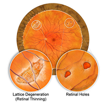
The retina is specialized nerve tissue layer lining in the inner surface of the eye that senses light and helps you to see. The majority of the retina's surface area is considered peripheral retina. The peripheral retina is devoted to side vision, also known as peripheral vision. Some people have areas of thinning in the peripheral retina, known as lattice degeneration. The thinning can be severe enough that a retinal hole can develop. it is more common in myopic eyes.
The cause is unknown but it is more common in people who are near-sighted (myopic) and tends to run in families
Usually, retinal holes and lattice degeneration cause no visual symptoms and are diagnosed on a routine eye examination. If retina has tear or holes, it can separate from the back wall of the eye. This is called retinal detachment. This can cause you to lose sight suddenly.
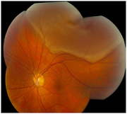
Retinal Detachment
Early detection and treatment of a retinal tear may help to prevent the formation of a retinal detachment or the expansion of a pre-existing detachment. It can help reduce the risk of vision loss associated with larger or more central detachment requiring a complicated retinal surgery.
Treatment of lattice is typically prophylactic. A “barrier” laser is applied to “tack down” the retina surrounding these lesions to avoid the possibility of retinal detachment. The laser is applied around the retinal tear or hole. Laser helps bond the retina to the wall of the eye, preventing a retinal detachment.
There is no risk of infection from the laser light.
Laser surgery can be performed in an outpatient setting allowing you to go home shortly after the procedure is finished.
The surgeon has great precision and control
Treatment is an outpatient procedure admission to hospital is not required. The patient sits at a slit lamp similar to that used in routine ophthalmic examination but modified to accept a laser fibreoptic cable. Drops are used to anaesthetize the cornea and a therapeutic contact lens is applied. As treatment is started, the patient experiences bright flashes of light but no pain. The eye must be kept as still as possible during treatment to ensure accurate application of the laser and to avoid damage to the fovea, a typical treatment takes about ten minutes.
Patient can communicate with the ophthalmologist in case of any discomfort so that the treatment can be safely interrupted. Commonly the patient is very dazzled after the treatment. No special precautions need to be taken after the laser. Additional eye drops or oral
therapy may be required in certain needed. Patient can resume daily routine soon after the therapy.